Example of a unilobar kidney (no slide)

Kidneys of small animals such as the rabbit and cat consist of a single lobe. Larger animals require more functional tissue than can be placed in a single lobe and thus have multilobar kidneys. This image of a unilobar kidney demonstrates the basic organization of cortical and medullary regions. The arcuate arteries (A) mark the junction between the cortex (C) and medulla (M). The outer cortex is covered by the capsule. The calyx (X) is a recess of the renal pelvis where urine collects. The lumen of the calyx is the clear space between the papilla of the pyramid and the ureter. Except for the calyx region, the medulla of a unilobar kidney is surrounded by cortical tissue.
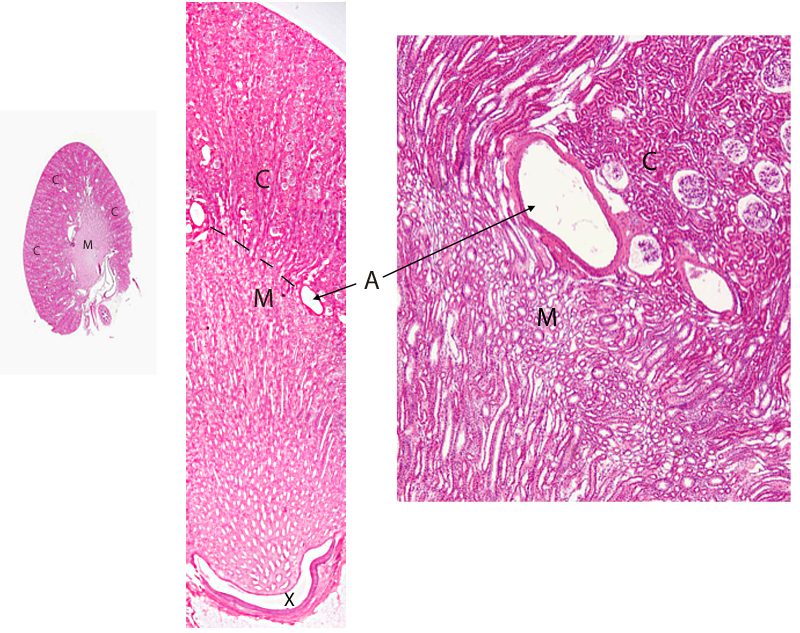
Three maginifications of the corticomedullary region of a unilobar kidney. H&E stain.